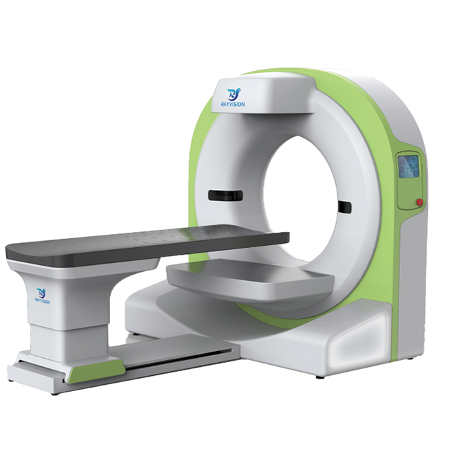
Vetease 3 Veterinary CT Scanning Imaging System is a multifunctional clinical diagnostic imaging solution designed specifically for veterinary clinics and emergency vet scenarios. With advanced 3D reconstruction capabilities, ultra-thin 0.14mm slice thickness, and proprietary CT-AIR imaging technology that effectively reduces motion artifacts, it ensures accurate and efficient imaging results even in critical situations. Compared to MRI, its compact design and faster scanning make it a practical choice in the CT scan vs MRI debate, particularly when time and motion sensitivity are crucial in small animal diagnostics. Lightweight, cost-effective, and clinically versatile.
CT imaging
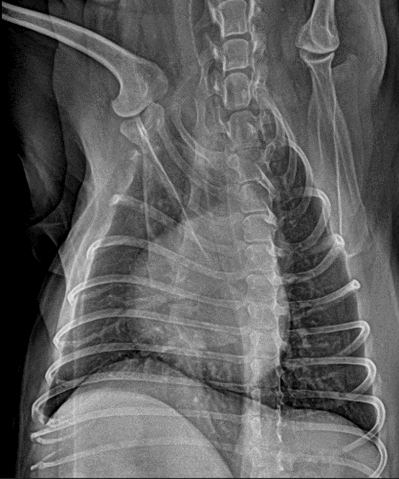
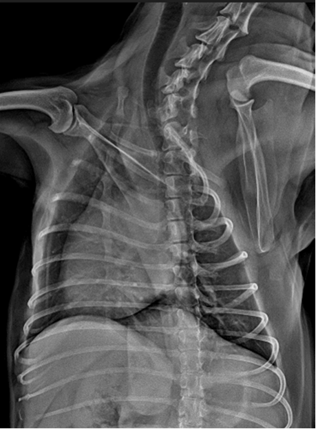
Vetease 3 CT image single-circle 600 image scanning slices, with a slice thickness of up to 0.14 mm, can display subtle anatomical details, provide veterinarians with clear and unambiguous image imaging reports, quickly locate the patient’s lesions, and make clinical decisions.
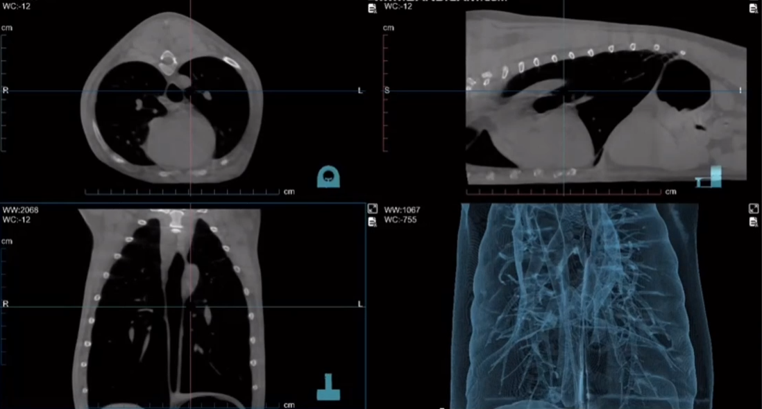
Vetease 3 CT Image post-processing technology can display lung structure more clearly, provide veterinarians with confident decision-making for the diagnosis of interstitial lung disease, pulmonary sarcoidosis, emphysema and other diseases in animal patients, and is sensitive to active lesions such as active tuberculosis and fungal infections, helping to distinguish active lesions from old scars, and provide a basis for the evaluation of early lesions in pets/precise staging and treatment planning.
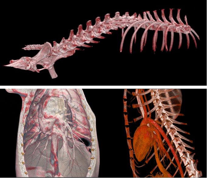
Vetease 3 has an ultra-high resolution of 0.14mm, which can clearly show the fine structure of the spine, small animal osteophytes, vertebral edge sclerosis, and articular process hyperplasia. It can perform multi-plane reconstruction and 3D imaging to help doctors accurately evaluate the relationship between the hyperplastic site and the surrounding structures (nerve roots, spinal canal).
Vetease 3 has an ultra-low radiation dose (about one percent of traditional spiral CT), which is safer for pets that need multiple reexaminations (such as elderly dogs and cats).
Evaluation of complex spinal lesions: When hyperplasia is combined with intervertebral disc calcification and vertebral compression fractures, bone lesions and calcified tissues can be distinguished.
Preoperative planning: For example, before spinal surgery (laminectomy, osteophyte removal), the scope of the lesion can be accurately located.
Advantages of small pets: The device is small in size and suitable for small and medium-sized animals such as dogs and cats, and has better local scanning effects on the cranial cervical and thoracic lumbar spine.
Dynamic evaluation of efficacy: The low-dose characteristics support regular reexamination to monitor the progression of hyperplasia or postoperative recovery.
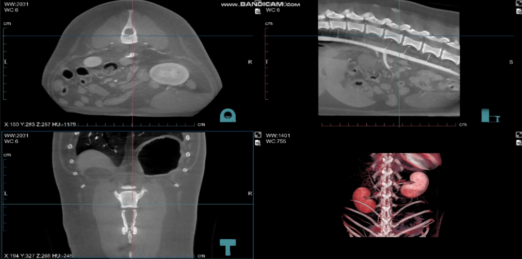
Vetease 3 CT generates high-quality 3D reconstruction images to help veterinarians fully evaluate the morphology, location, structure and possible lesions of the kidney.
Kidney stones: can clearly show the location, morphology and relationship of stones to other kidney structures.
Kidney tumors: Vetease 3 CT’s high-resolution images can help determine the size, boundaries and invasion of adjacent structures of the tumor.
Cysts and other structural abnormalities: Vetease 3 CT can provide high-contrast images, and small cysts or other structural abnormalities can be clearly displayed.
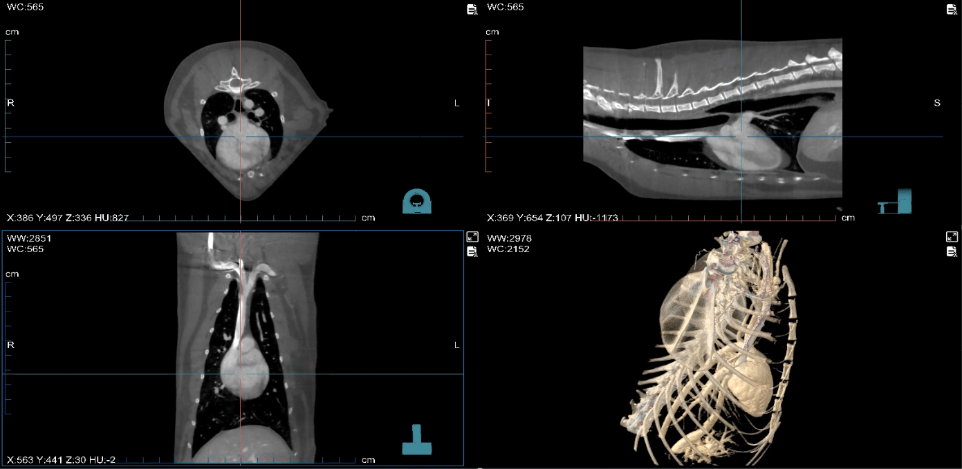
Vetease 3 can clearly display the heart structure and pathological changes of small animals, such as valvular disease, ventricular enlargement, cardiac tumors, etc.
At the same time, the three-dimensional reconstruction image of the heart facilitates a comprehensive assessment of the size, shape, blood vessels and valve status of the animal’s heart. It helps to identify complex heart lesions, especially accurate diagnosis and treatment plans for valvular heart disease and congenital heart defects.
Combined with contrast agents for cardiac angiography, it enhances the imaging effect of the heart and blood vessels and helps identify problems such as vascular stenosis, aneurysms, and poor blood flow. Helps veterinarians diagnose coronary heart disease, cardiovascular abnormalities and circulation conditions in pets and small animals
Section display: Display teeth and surrounding structures on the coronal, sagittal and horizontal planes, clearly showing the state of the root, crown and root of the tooth and changes with the surrounding bones. Identify periapical inflammation, apical lesions, etc.
Surface reconstruction images; veterinarians can comprehensively evaluate the morphological changes of teeth and the extent of caries, such as surface lesions, tooth wear, caries, periodontal disease, etc.
Precisely locate the lesion area: VR reconstruction technology provides a complete three-dimensional perspective and accurately locates the lesion area, providing strong clinical evidence for the diagnosis of complex bone, joint or visceral diseases in small animals. Veterinarians can locate the lesion more accurately and develop effective treatment plans.
Dynamic and Static DR imaging
Rapid scanning can be completed in as little as 12S, with a spatial resolution of ≥3.0lp/mm and a wide field of view, providing clear imaging.
Animal-specific image algorithm software
Our R&D team cooperated with top medical schools Southern Medical University and China Agricultural University to jointly develop imaging post-processing technology specifically for animals and applied it clinically, achieving satisfactory results.