Basic information: Cat, aged nearly 2 years, female already spayed
Symptoms: The cat presented with dry heaving, panting, hunger strike and decreased stool volume
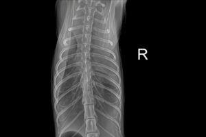
Initial diagnosis: Temperature 40°C, DR image shows large pleural effusion

Diagnostic thoughts:
1. Common causes of pleural effusion in cats are cardiac disease (e.g. hypertrophic cardiomyopathy), tumours (e.g. lymphoma or thymoma), infections (e.g. FIP), and coeliac disease. In addition, there may be other causes such as diaphragmatic hernia or hypoproteinaemia, but they are less common.
2. In affected cats, an elevated body temperature of up to 40°C suggests that infection or inflammation may be present. FIP usually causes fever as well, especially in wet FIP where pleural effusion is common. However, the definitive diagnosis of FIP is complex and requires a combination of other tests. In addition, although cardiac disease does not always cause fever, cardiogenic effusion may be accompanied by other symptoms, such as shortness of breath, and this cat did have wheezing.
3. DR showing a large pleural effusion should be followed by thoracentesis and analysis of the effusion, including cytology, protein levels, Levantine test, bacterial culture and PCR testing. This helps to differentiate between leaking fluid, oozing fluid or coeliac fluid. If the fluid is coeliac, it may point to a lymphovascular abnormality or heart disease; if it is exudative, infection or tumour may need to be considered.
4. Ultrasound of the heart is necessary to assess the structure and function of the heart and to rule out hypertrophic cardiomyopathy or other heart disease. In addition, abdominal ultrasound may help to detect underlying tumours or organ abnormalities such as hypoproteinaemia due to liver or kidney problems.
5. Routine blood and biochemical tests are also necessary. Hyperglobulinaemia may indicate FIP, while low albumin may point to other problems. Special attention needs to be paid to the total protein to albumin ratio. an SAA test can help determine the degree of inflammation.
6. the sequence of tests needs to be optimised given that the pet owner is looking for cost savings.
Diagnostic protocol flow:
I. Primary Emergency Treatment
Stabilise vital signs:
Immediate thoracentesis drainage to relieve respiratory distress;
Split fluid accumulation into EDTA tubes (cytology) & sterile tubes (biochemistry/PCR)
| Test items | Clinical significance |
| 1. Pleural fluid analysis | |
| Cytological classification | Identification of tumour cells/purulent exudate (bacterial infection) |
| Levantine test + protein quantification | Determination of leaky fluid exudate celiac disease |
| FCoVPCR | Exclusion of feline infectious peritonitis (FIP) |
| 2. Blood routine + 12 items of biochemistry | |
| SAA, Globulin | Assessment of inflammation levels (FIP is often associated with hyperglobulinaemia) |
| ALB/GLOB ratio | ratio < 0.4 highly suggestive of FIP |
| 3. Cardiac ultrasound | Screening for structural heart disease such as hypertrophic cardiomyopathy (HCM) |
III. Etiological Prioritisation and Response
Based on test results, dispose of them in the following probability order:
1. Highly suspicious: FIP (wet)
Supporting evidence: yellowish fluid, Livanta positive, high globulin, ALB/GLOB ratio <0.4;
Rapid confirmation of diagnosis: send for PCR of the mutated locus;
2. Secondary: cardiac disease (cardiogenic effusion)
Supporting evidence: cardiac ultrasound showing left ventricular wall thickness >6mm and left atrial dilatation;
Confirmatory means: ProBNP test (>500 pmol/L);
3. Other possibilities: tumour/bacterial pyothorax
Supporting evidence: heterogeneous cells/bacteria seen on cytology;

Hedgehog uterine effusion – ultrasound clinical examination
Uterine pus accumulation in hedgehogs may be accompanied by other symptoms such as loss of...
Per saperne di più
Emoplasmosi felina senza protocolli di analisi biochimica
Il micoplasma distrugge le membrane degli eritrociti → emolisi intra ed extravascolare → emolisi secondaria immuno-mediata (IMHA) →...
Per saperne di più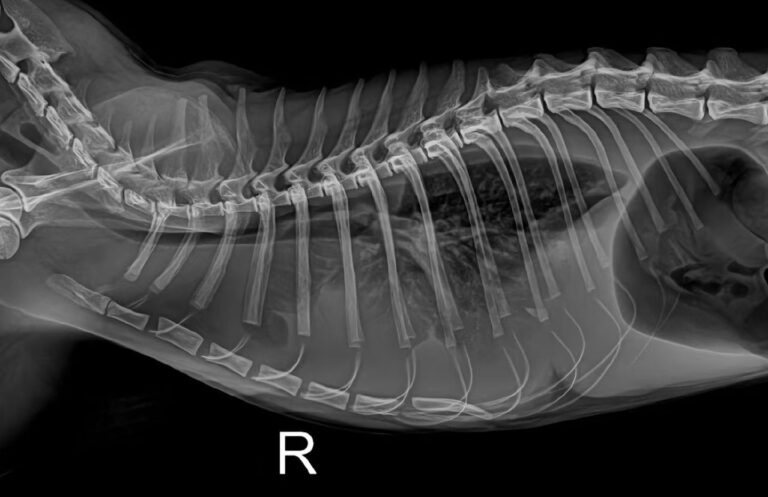
Caso di versamento pleurico in un gatto: procedura di diagnostica d'urgenza
Le cause più comuni di versamento pleurico nei gatti sono le malattie cardiache (ad esempio, la cardiomiopatia ipertrofica), i tumori (ad esempio, ....
Per saperne di più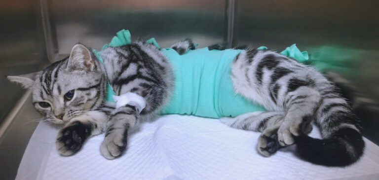
Perché sterilizzare i gatti? Tempistica ottimale e cure post-chirurgiche
La sterilizzazione è una responsabilità fondamentale per i proprietari di animali domestici, in quanto migliora la salute dei felini e riduce il randagismo...
Per saperne di più